






Surgical removal of skin lesions
A precise, safe and professional solution for your skin.
Skin formations such as moles, growths, cysts, lipomas or suspicious pigment changes often require professional examination and sometimes surgical removal. At our dermatology clinic, we offer professional procedures aimed not only at aesthetics, but also at preventing more serious diseases, such as skin cancer.
When is surgical removal necessary?
We recommend surgery especially in the case of:
- suspicious or changed pigmented nevi (birthmarks)
- fast-growing or painful growths
- subcutaneous formations (lipomas, cysts, atheromas)
- chronically irritated growths in the area rubbing against clothing
- unaesthetic formations with the recommendation of their histological examination
How is the procedure performed?
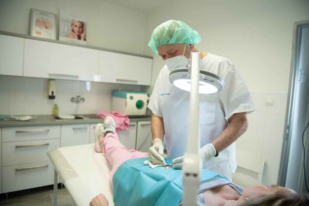
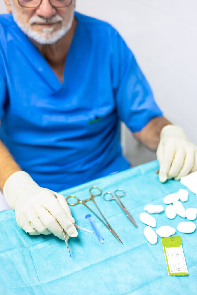
Surgical removal is performed on an outpatient basis under local anesthesia. After anesthesia, the formation is excised with a scalpel or electrosurgically. The wound is then treated and sutured (if necessary). If necessary, the removed material is sent for histological examination to accurately determine its nature.
The procedure itself usually takes 20-40 minutes. The patient goes home without the need for hospitalization. We are extremely consistent in adhering to hygiene standards and use modern surgical instruments and gentle techniques.
Postoperative care
After the procedure, it is important to follow the doctor's recommendations:
- keep the wound clean and protected
- avoid excessive movement in the area of the procedure
- undergo a check-up and removal of stitches (usually 7–14 days after the procedure)
- protect the area from the sun (to minimize the risk of scarring)
Why choose us?
- experienced team of doctors
- individual approach and thorough diagnostics
- use of gentle and precise techniques
- aesthetic suturing and scar minimization
- quick return to normal activities
Removal of unwanted skin growths
fibromas, keratoses, papillomatous formations, hemangiomas
Pigmented nevi and other benign skin lesions
The most common benign skin lesions include:
- pigmented nevi/ nevus cellular, intradermal, junctional, confluent, blue nevus, Spitz nevus, Halo nevus…./
- organoid nerves / naevus telangiectaticus, angiomatous nevus, lipomatous naevus, etc.
- seborrhoeic and actinic keratoses, hidradenomas,
- fibroids – soft, hard
- haemangiomas – capillary, cavernous
- lipomas , chondromas
- epidermoid cysts
- atheromas
- histiocytomas
- pyogenic granuloma
Benign/non-malignant/tumors Skin and pigmented nevi usually do not require treatment, the exception being if they are located in areas of chronic irritation or if there is a possibility of degeneration into a malignant tumor.
Treatment options – total surgical excision with histological examination
– electrocoagulation
– laser therapy
– cryotherapy


Malignant skin tumors
Malignant melanoma
Malignant melanoma is a highly malignant neoplasm characterized by rapid spread of metastases by hematogenous and lymphogenous routes
It arises from melanoma cells. The cause of its development is unknown, but it is thought to be genetic predisposition, viral etiology and the impact of excessive UV radiation.
Occurs most often in white people.
It can occur anywhere on the skin, most commonly hairy part of the head, face, limbs.
It most often forms from pigmented nevi or appears on previously unchanged skin.
Clinical picture – dark brown to black formation, at the skin level or a nodule to a bump, oozing, bleeding, subjective itching to pain. Characteristic of malignant melanoma is rapid growth of the formation.
There is also an amelanotic type of melanoma – it mimics moles, verrucas, and fibromas.
Prevention – it is important to pay attention to pigmented nevi, early recognition of dysplastic nevi and their subsequent early removal with histological examination
Treatment – urgently send to the melanoma committee where the diagnostic and therapeutic procedure will be determined.


Squamous cell carcinoma
it is malignant tumor which begins as an inconspicuous keratosis of a yellow-brown color with a rough surface, grows relatively quickly and turns into a bump with a cauliflower-like appearance, often bleeds and destroys the surrounding tissue and metastases occur in the regional lymph nodes and can gradually spread to various organs.
It is most often localized to face – forehead, ears, lips, tongue.
The cause of the occurrence is stated genetic predisposition, influence of UV radiation, ecological factors/tar, tobacco, petroleum products/
Prevention and treatment – early diagnosis and early surgical excision of the tumor are important


Basal cell carcinoma
is most common malignant tumor, occurs in older people.
The most important pathogenetic factor is actinic radiation, especially the UV-B component, occurs most often in areas with intense sunlight.
The preferred location of basal cell carcinoma is exposed parts of the skin /face, neck, nape, ears and lower legs/
The tumor begins inconspicuously as a small, pinkish-red elevation of the skin, gradually growing into a brownish nodule with a pearly surface. Small telangiectasias, scabs, bleeding, and oozing are common.
The neoplasm grows relatively slowly and does not metastasize. It grows locally and destroys only the surrounding tissue.
Prevention and treatment – prevent excessive exposure to UV radiation, early diagnosis and surgical excision with histological confirmation of the diagnosis. It is also possible to use cryotherapy, electrocoagulation or application of local cytostatics.

